Pillar II · AI HealthAI-powered automation of
AI-powered automation of
MS radiological follow-up.
An AI deep-learning pipeline that auto-segments white matter demyelinating plaques on 3D volumetric brain MRI and color-codes longitudinal lesion changes: turning time-consuming manual review into objective, reproducible results.
The Problem
In Multiple Sclerosis follow-up, radiologists manually cross-reference every lesion across longitudinal cranial MRI scans. The process is highly time-consuming (up to 45 minutes per study) and introduces significant user-dependent variance into disease monitoring.
The Solution
Our deep-learning pipeline auto-segments white matter demyelinating plaques on 3D volumetric brain MRI, then color-codes lesions as stable, new (interval), progressed, or regressed when comparing current scans with prior studies: delivering objective, reproducible metrics automatically. The same longitudinal change-detection core extends to adjacent AI health applications, including surgical decision support, as the platform matures.
Interactive Demo
Before & after AI segmentation.
Drag the slider to compare a baseline cranial MRI with the AI-segmented follow-up scan. Each lesion is automatically color-coded by longitudinal change status.
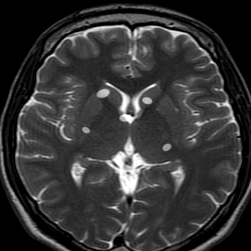
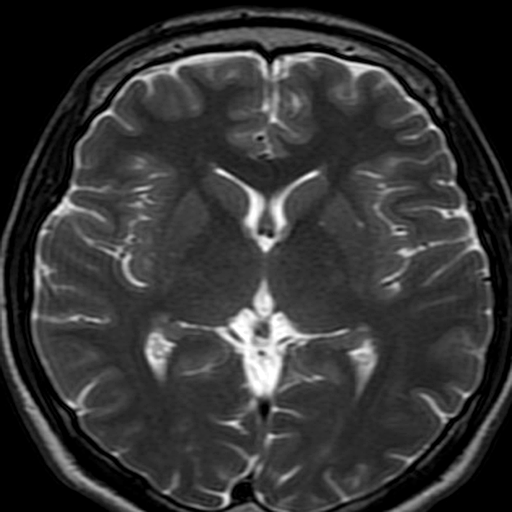
Baseline
Follow-up · AI Segmented
AI lesion differential:StableNew (Interval)ProgressedRegressed
Reference MRI: Wikimedia Commons (CC BY-SA 4.0).
Simulated illustration of 3D volumetric segmentation output for demonstration purposes.
Clinical Workflow
From MRI scan to clinical insight.
Our end-to-end pipeline integrates natively with hospital PACS/HIMS systems, turning raw MRI volumes into color-coded longitudinal insights automatically.
Stage 1 / 5
DICOM Ingest
Cranial MRI volumes ingested via DICOM parser from PACS.
Native PACS/HIMS query & storage protocols.
Integrates with:PACSHIMSDICOMTeletıpe-NabızREST API
Core Technological Pillars
Engineered for clinical precision.
3D Spatial Context
Unlike 2D slice-by-slice models, our model leverages full volumetric context, leveraging spatial continuity across slices to estimate plaque volumes accurately.
Differential Coding
Algorithms compare co-registered longitudinal volumes, highlighting stable, new, progressed, and regressed lesions under a unified color scheme.
Interoperability
Engineered to interface natively with PACS, HIMS, and state e-health portals like Teletıp and e-Nabız using secure REST APIs.
NEDA-3 Imaging Component
The MRI half of The MRI half of NEDA-3, automated.
Treatment decisions hinge on one question: is the disease quiet? The imaging half of NEDA-3: new or enlarging T2 lesions and Gd-enhancing lesions: is exactly what our registered follow-up pipeline detects automatically.
New / enlarging T2 lesion
FLAIR pair registration
Gd-enhancing lesion (active disease)
T1 post-contrast (roadmap)
Clinical relapse
EDSS / neurologist
Disability worsening
EDSS
2 of 4 components automated today: the two that are fully objective.
Interactive Simulator
Run the pipeline step by step.
Experience how Cordistronic's AI processes a cranial MRI volume: from DICOM ingestion to color-coded longitudinal output.
T2 AXIAL · RAW
Idle
Step 0
Upload Brain MRI
Select a cranial MRI volume to process through the AI pipeline.
Step 1
DICOM Ingestion
Step 2
AI Segmentation
Step 3
Differential Coding
Step 4
Color-Coded Result
Clinical Trial Enrollment
Join the clinical validation network.
We're recruiting hospital partners for multi-center clinical validation of our AI MS radiology pipeline. Express interest below.
Multi-Center Validation
Join the clinical validation network across Türkiye & EU.
Early Access
Pilot the AI pipeline before CE/MDR clearance.
Co-Authorship
Contribute to clinical evidence and academic publications.
TRL 5 → TRL 7 pathway. Clinical validation is a required step for CE/MDR class-IIa clearance. Your participation directly accelerates deployment.
Clinical Evidence
Before & after patient outcomes.
Illustrative case studies demonstrating how Cordistronic's solutions transform clinical workflows: from subjective, time-consuming processes to objective, reproducible precision.
Longitudinal MS Follow-Up: 18-Month Tracking
34-year-old female · RRMS diagnosis · 3 cranial MRIs over 18 months
A relapsing-remitting MS patient monitored across three longitudinal MRI scans. The AI pipeline detected disease activity changes that manual review had flagged inconsistently.
Analysis method
Manual review
Deep-learning segmentation
AutomatedLesion detection consistency
Operator-dependent
fully reproducible
ObjectiveInterval change detection
Qualitative estimate
Color-coded (4 categories)
QuantitativeBoundary precision
Variable
Maximized
OptimizedClinical narrative
1The patient presented with 12 established white matter lesions at baseline. Manual cross-referencing across subsequent scans took approximately 45 minutes per study and produced inconsistent boundary delineation between reviewers.
2Our deep-learning pipeline processed each 3D volumetric brain MRI automatically, segmenting all lesions and comparing them against the baseline with full volumetric context.
3At month 12, the pipeline flagged 2 new interval lesions (red) and 1 progressed lesion (amber): changes that manual review had noted but struggled to quantify precisely.
4By month 18, 1 lesion showed regression (green). The color-coded output enabled the neuroradiologist to deliver an objective, reproducible report automatically: transforming the clinical follow-up workflow.
Case studies are illustrative and based on aggregated clinical scenarios. Actual patient data is confidential and processed under institutional review board oversight.
Interested in clinical pilot integration?
We're seeking hospital partners for PACS/HIMS integration, multi-center clinical validation, and CE/MDR regulatory pathway.